Visibly Complex
 Tom
Glaser, Associate Professor,
Department of Internal Medicine, University of Michigan
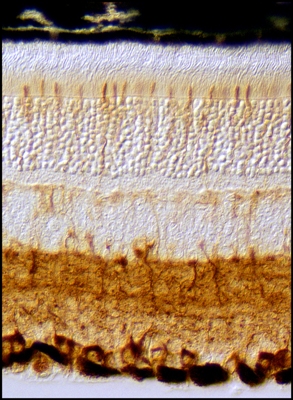
This retina of a laboratory mouse is viewed with Nomarski optics
and stained
with an antibody to identify ganglion cells, cone photoreceptors
and the inner
plexiform layer (brown). A quarter of a millimeter across, this
layered tissue
covers the inside surface of the eye. The human retina is
similar, and functions
as a highly organized switchboard that records, processes and
conveys all visual
information from the outside world to the brain. In this image,
light first passes
through the lens and then enters the retina to excite the cone
and finger-like rod
photoreceptors located at the back of the eye near the pigmented
epithelial layer
(black). The resulting electrical signals are then relayed
stepwise, via delicate
neural tendrils, to the ganglion cells, whose projections join
to form the optic
nerve and travel to the brain.
|
 Fiber
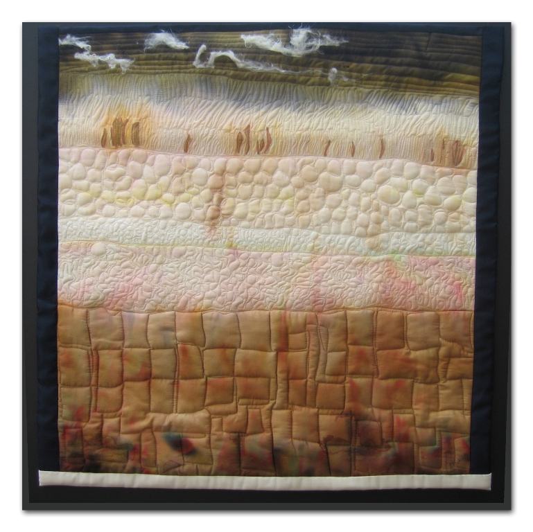
Artists @ Loose Ends encourages members to explore new ideas and
techniques, inspires and nurtures creativity. By sharing our work
in private and public venues, we express our passion for the textile
medium.
Fiber
Artists @ Loose Ends encourages members to explore new ideas and
techniques, inspires and nurtures creativity. By sharing our work
in private and public venues, we express our passion for the textile
medium.