Gutsy
 Blair Madison, Ph.D., Postdoctoral Fellow, and Andrea Waite, Blair Madison, Ph.D., Postdoctoral Fellow, and Andrea Waite,
Graduate Student, Cell & Developmental Biology, University of
Michigan
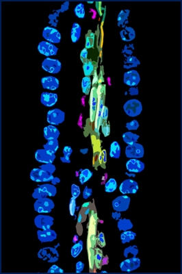
The surface of the intestine is lined by millions of finger-like
structures (villi) that
extend into the lumen of the intestine to provide enormous
surface area for
absorption of nutrients. This photomicrograph shows a portion of
an intestinal
villus. The blue ovals are nuclei of individual cells that cover
the villus’ surface.
These cells absorb proteins, sugars and fats from food in the
gut lumen. The
absorbed nutrients are processed by these cells, and then
secreted into blood
vessels that spiral up into the center of the villi (pink). The
muscles (beige) in the
center squeeze the villi to pump nutrients into the main blood
stream.
|
Donna DeSoto
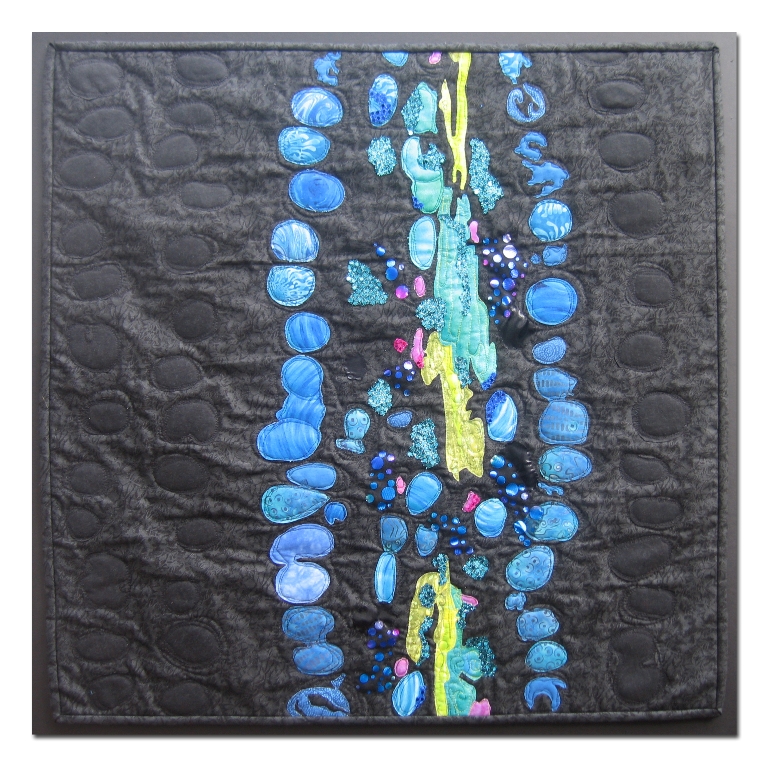
Two things initially drew me to this photo: the beautiful blue
colors and the organic shapes! I have an extensive collection
of all shades of blue fabric, and I was eager to use it, set off
against a stark black background. This piece was constructed
with commercial cotton fabric, some enhanced by the addition
of paint and/or stamping. Raw edged shapes were applied
to the background by free motion quilting. This quilt was
made in honor of my father-in-law, Oscar DeSoto, who was
born in Cuba.
Back to Gallery
|
 Fiber
Artists @ Loose Ends encourages members to explore new ideas and
techniques, inspires and nurtures creativity. By sharing our work
in private and public venues, we express our passion for the textile
medium.
Fiber
Artists @ Loose Ends encourages members to explore new ideas and
techniques, inspires and nurtures creativity. By sharing our work
in private and public venues, we express our passion for the textile
medium.